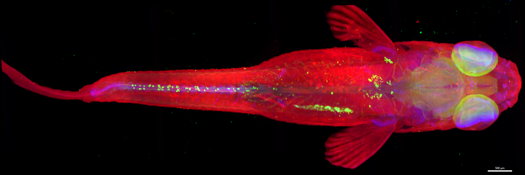
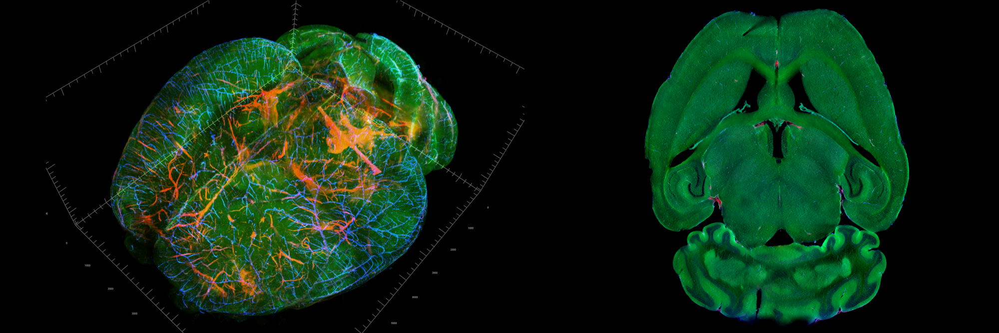
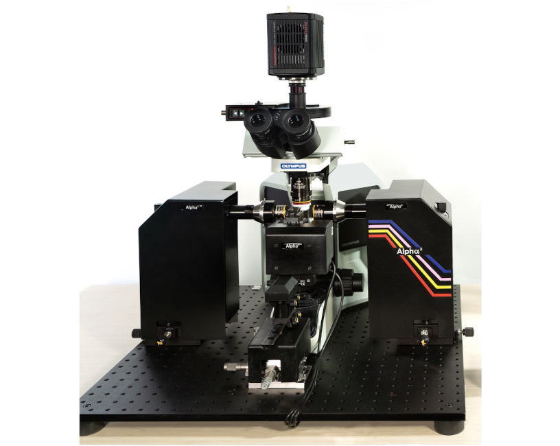
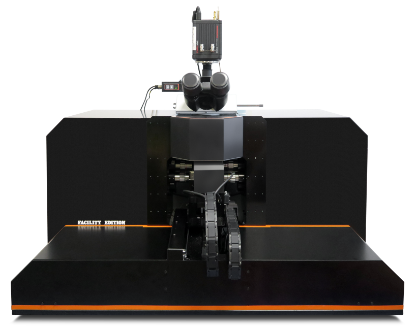
Light Sheet Microscopy


Ultra Thin Light Sheet
Cleared Tissue & Live Specimen
Beyond Light Sheet Imaging
Optimal Image Quality
Ultra thin sectioning for sub-cellular resolution

Patented high-illumination NA and focus sweeping eliminate trade-offs between resolution and field of view.
Conventional Light Sheet Fluorescent Microscopy


Trade-off between resolution and the field of view.
Ultra thin sectioning for sub-cellular resolution

Conventional Light Sheet Fluorescent Microscopy

PEGASOS cleared whole mouse brain. Courtesy of Pr. Edna Hardeman and Bio-Imaging Facility (BMIF) of UNSW Sydney.
Optimal Image Quality
Maximized resolution across the whole field of view

Patented real time focus sweeping technology.
Conventional Light Sheet Fluorescent Microscopy

Gaussian beam divergence.
Ultra thin sectioning for sub-cellular resolution

Conventional Light Sheet Fluorescent Microscopy

PEGASOS cleared whole mouse brain. Courtesy of Dr. Genaro Hernandez, University of Texas Southwestern, Dallas, USA
Optimal Image Quality
Maximum light coverage and intensity homogeneity
Simultaneous dual illumination with high lateral NA provides optimal sample penetration, even with thick samples, and efficient artifact removal when light is absorbed or scattered. This state-of-the-art technology enables true qualitative and quantitative imaging.

Patented Simultaneous dual illumination and high lateral NA illumination technologies.
Conventional Light Sheet Fluorescent Microscopy
Lack of intensity homogeneity, shadow or stripe artifacts are common in light sheet microscopy despite more or less complex optical solutions to eliminate undesirable effects associated with light sheet fluorescence microscopy.

Optical architecture results in insufficient light penetration and poor image quality due to unwanted artifacts and shadows.
Flexibility and Modularity
Fast and easy sample mounting solutions
All mounting accessories are conveniently adapted for all sample types and sizes. Sample integrity and physiological conditions are maintained during image acquisition. Mounting accessories and sample chamber are highly resistant to corrosive media and can be easily cleaned.

Small Sample Mounting
Large Sample Mounting
Flexibility and Modularity
Multi-sample acquisition
Light sheet microscopes are well suited for high throughput imaging experiments, offering an extended sample holder with clamps to accommodate multiple samples. Software resources handle the acquisition of individual stacks under the desired imaging conditions.

Examples of sample mounting on sample holder.
Flexibility and Modularity
Configurable microscopy systems with a wide range of illumination and detection lenses
Alpha3 Facility Edition is a fully automated system that performs microscope configuration changes and automatic adjustments when changing objective magnification for both illumination and detection objectives.

Compatibility between illumination and detection objectives.
Unrivaled User Experience
Automated multi-scale microscopy systems
Alpha3 Facility Edition is a fully automated system that performs microscope configuration changes and automatic adjustments when changing objective magnification for both illumination and detection objectives.
The user can begin an imaging session with rapid sample screening using a low-magnification objective, followed by image acquisition using a higher-magnification objective on a single or multiple regions of interest.
After the user selects the desired region of interest, the system automatically selects illumination and detection objectives.
Unrivaled User Experience
3D viewer with interactive volume scan settings
The LINDA interface extension is at the heart of the user experience, generating a 3D volume preview on the fly to explore the sample before scanning. The wizard provides a live view of the current slice and “builds” a 3D rendering as the user explores the sample. LINDA allows the user to interact with the 3D rendering to set the scan volume and acquisition parameters.

3D view of a sample with interactive volume scan settings.
Unrivaled User Experience
Imaging PaaS is a free virtual bio-imaging platform that provides on-demand services. You can access expert support for your clearing protocols, powerful computing resources for your image analysis, or advanced microscopy solutions at your convenience.