Applications
Physiology / Whole organs
Whole Mouse Brain
PEGASOS cleared whole mouse brain, Life-act EGFP transgenic actin filaments staining, tdTomato transgenic MARCKS plasma membrane staining. Objective: 4X/0.28NA/30WD.
Courtesy of Pr. Edna Hardeman, Bio-Imaging Facility (BMIF) of UNSW Sydney.
Visualized with Imaris Software
Human Gastric Organoids
Human gastric organoids in PBS1X, with nuclear DAPI staining and membrane CDH1/Alexa546 Staining. Objective: 40X/0.80NA/3.3WD.
Courtesy of Elena Amendola, Monte S. Angelo University Federico II.
Visualized with Imaris Software
Zebrafish Larva, Multi-Scale Imaging
CUBIC R+ (RI 1.52) cleared whole zebrafish larva (10.5 x 3 x 2.2 mm), YOYO-1 nuclear staining, DiD lipophilic staining, Alexa 647 anti-TH dopaminergic neurons staining. Large Scale Imaging, illumination objective: 4X (UPLFLN4X), detection objective: 4X (XLFLUOR4X/340). Small-Scale Imaging, illumination objective: 10X, detection objective: 10X.
Courtesy of Matthieu Simion, TEFOR Paris-Saclay.
Visualized with Imaris Software
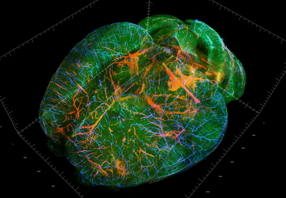
Pig Brain, Multi-Scale Imaging
iDisco cleared 2×2 stitched pig brain imaging at 638nm illumination, emission filter at 690/50 nm. Large Scale Imaging, illumination objective: 4X (UPLFLN4X), detection objective: 4X (XLFLUOR4X/340). Small-Scale Imaging, illumination objective: 10X (LMPLFLN10X), detection objective: 10X (XLPLN10XSVMP-2).
Courtesy of Dr. Alexander Rapp, TU Darmstadt, Germany.
Visualized with Imaris Software
Cleared Brain Tissue
CCK+interneurons in the entorhino-hippocampal formation in the mouse brain (GFP) and Wfs1-marked pyramidal. Objective: Olympus XLFLUOR4X/340 (4X/0.28NA/30WD).
Courtesy of Nóra Henn-Mike, Institute of Physiology, Medical School, University of Pécs.
Visualized with Imaris Software
Zebrafish Brain
Cleared whole zebrafish brain, DAPI staining, Alexa 488 neuronal staining, DiD neuronal staining. Objective: Olympus XLPLN10XSVMP-2 MPE (10X/0.60NA/8WD).
Courtesy of Isabelle Robineau & Matthieu Simion, TEFOR Paris-Saclay.
Visualized with Imaris Software
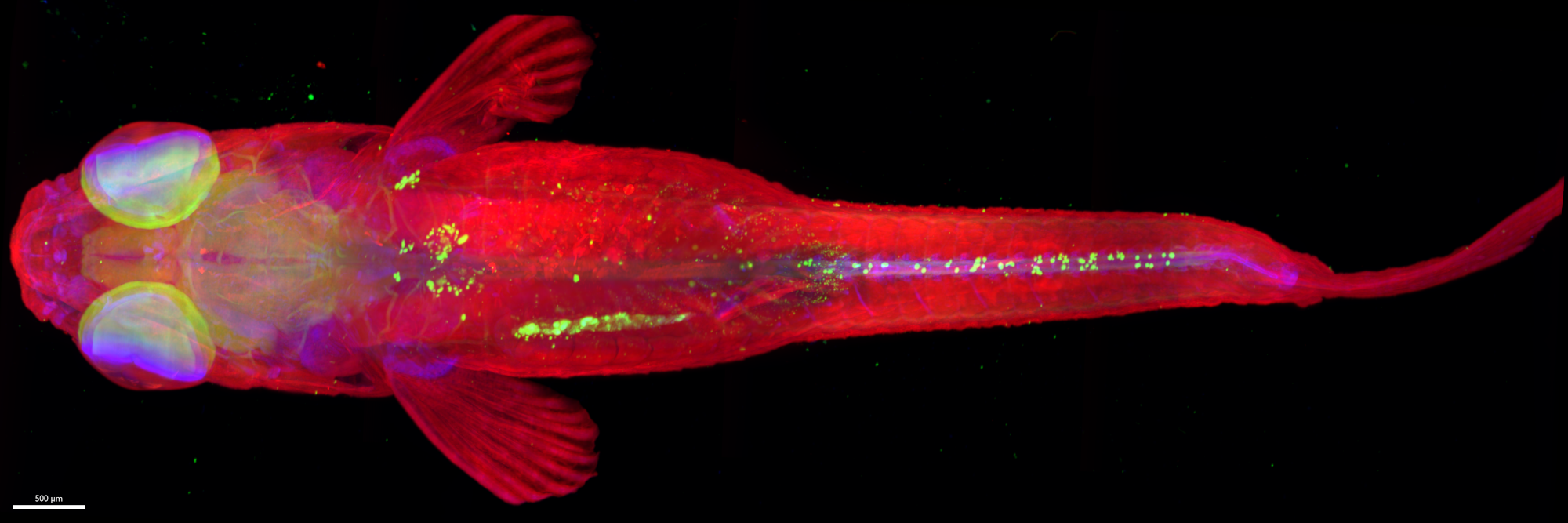
CUBIC R+ cleared whole Zebrafish Larva
CUBIC R+ cleared whole Zebrafish Larva, YOYO-1 nuclear staining, DiD lipophilic staining, Alexa 647 anti-TH dopaminergic neurons staining. Objective, UPLFLN4X (4X/0.28NA/30WD).
Courtesy of Dr. Matthieu Simion (TEFOR Paris-Saclay).
Visualized with Imaris Software

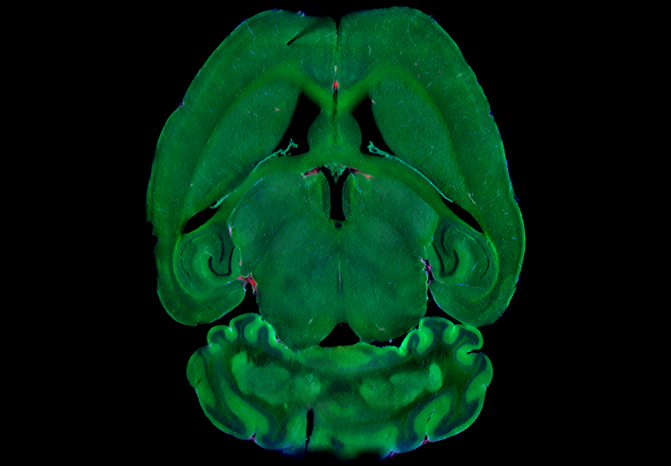
Whole mouse brain


PEGASOS cleared whole mouse brain, Life-act EGFP transgenic actin filaments staining, tdTomato transgenic MARCKS, plasma membrane staining. Objective, UPLFLN4X (4X/0.28NA/30WD).
Courtesy of Pr. Edna Hardeman and Bio-Imaging Facility (BMIF) of UNSW Sydney.
Visualized with Imaris Software
Whole mouse brain
PEGASOS cleared whole mouse brain, Life-act EGFP transgenic actin filaments staining, tdTomato transgenic MARCKS, plasma membrane staining. Objective, UPLFLN4X (4X/0.28NA/30WD).
Courtesy of Dr. Genaro Hernandez, University of Texas Southwestern, Dallas, USA.
Visualized with Imaris Software

Retina / Morphological analysis
Mouse Retina
Retinal whole mount from mouse with Thy1-GCAMP3. Objective: Olympus XLFLUOR4X/340 (4X/0.28NA/30WD).
Courtesy of Tamas Kovács-Öller, Szentágothai Research Centre (SzKK).
Visualized with Imaris Software
Physiology / Morphological analysis
Zebrafish Larva
CUBIC R+ cleared whole zebrafish larva, YOYO-1 nuclear staining, DiD lipophilic staining, Alexa 647 anti-TH dopaminergic neurons staining. Objective: Olympus XLSLPN25XGMP (25X/1.00NA/8WD).
Courtesy of Matthieu Simion, TEFOR Paris-Saclay.
Visualized with Imaris Software
Zebrafish Larva
Cleared whole zebrafish larva, Alexa 488 post-synaptic staining, Alexa 555 glial cells staining. Objective: Olympus XLPLN10XSVMP-2 MPE (10X/0.60NA/8WD).
Courtesy of Elodie Machado & Matthieu Simion / TEFOR Paris-Saclay.
Visualized with Imaris Software
Zebrafish Larva
CUBIC R+ cleared whole zebrafish larva, YOYO-1 nuclear staining, DiD lipophilic staining, Alexa 647 anti-TH dopaminergic neurons staining. Objective: Olympus XLFLUOR4X/340 (4X/0.28NA/30WD).
Courtesy of Matthieu Simion, TEFOR Paris-Saclay.
Visualized with Imaris Software
Zebrafish Larva
CUBIC R+ cleared whole zebrafish larva, YOYO-1 nuclear staining, DiD lipophilic staining, Alexa 647 anti-TH dopaminergic neurons staining. Objective: Olympus XLPLN10XSVMP-2 MPE (10X/0.6NA/8WD).
Courtesy of Matthieu Simion, TEFOR Paris-Saclay.
Visualized with Imaris Software
Multiscale Imaging of Adult Zebrafish
Multiscale Imaging of Adult Zebrafish. Objectives: 4X 0.28NA, 10X 0.6NA.
Courtesy of Matthieu Simion, TEFOR Paris-Saclay.
Visualized with Imaris Software
Axolotl
ECI-transparized Axolotl Imaging in two channels (RFP, far red) with 10X NA 0.6 objective.
Courtesy of Matthieu Simion, TEFOR Paris-Saclay.
Visualized with Imaris Software
Brain / Neuronal activation mapping
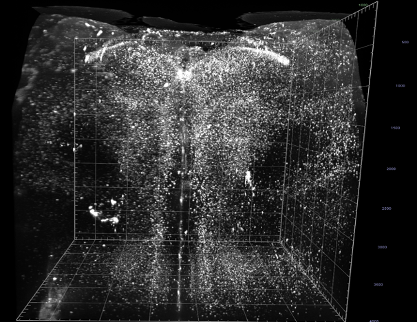
Cleared Mouse Brain
iDisco cleared brain with CFOS marking in green channel. Objective: Olympus UPLFLN4X (4X/0.28NA/30WD).
Courtesy of Dr. Genaro Hernandez, University of Texas Southwestern, Dallas, USA.
Visualized with Imaris Software
Zebrafish Brain Imaging
Adult Zebrafish Brain imaged by PhaseView Alpha3 Light Sheet Microscope, at 10X/NA0.6.
Courtesy of Matthieu Simion, TEFOR Paris-Saclay.
Visualized with Imaris Software
© 2021 PhaseView
Developmental Biology
Ganglion Imaging
Ganglion Imaging by PhaseView Alpha3 Light Sheet Microscope.
Visualized with Imaris Software
© 2016 PhaseView
Mouse Embryo Imaging
Mouse embryo in the E11.5 state transparized with CUBIC-L/R. Muscular system (red), nervous system (white).
Courtesy of Eglantine Heude and Frida Sanchez Garrido, Museum National d’Histoire Naturelle, France.
Visualized with Imaris Software
© 2024 PhaseView
Zebrafish Egg
15 hours-timelapse of a zebrafish egg with endothelial GFP staining and transmited light.
Courtesy of Beatriz Novoa & Antonio Figueras (Inmunology and Genomics Lab, Institute of Marine Research.
Visualized with Imaris Software
Astyanax Morpho Development Imaging
Astyanax Morpho Development, 28 hours time lapse, mCherry nuclei staining, GFP cytoplasmic staining in head.
Visualized with Imaris Software
© 2023 PhaseView
Drosophila Imaging
Drosophila fly egg, RFP nuclei staining, GFP membrane staining. Objective: 60X NA/1.0 water.
Visualized with Imaris Software
© 2023 PhaseView
Zebrafish Embryo Imaging
Zebrafish Embryo 36 hpf, RFP / GFP staining.
Visualized with Imaris Software
© 2023 PhaseView
Cell Biology
tumor spheroids
Imaging of tumor spheroids in far red.
Courtesy of Dr. Simona Mura, Institut Galien Paris-Saclay, France.
Visualized with Imaris Software
© 2023 PhaseView
Zebrafish Cellular Division
Imaging of Zebrafish Cellular Division, Nuclei NLS, H2B mCherry staining.
Courtesy of Nadine Peyrieras, BioEmergences, Gif sur Yvette, France.
Visualized with Imaris Software
© 2023 PhaseView
High Speed Imaging
Blood Cell Flow
Imaging of Blood Cell Flow in Zebrafish.
These rapid circulatory movements in live zebrafish were captured for only a few seconds.
Visualized with Imaris Software
© 2023 PhaseView
Red Core Imaging
This example of high-speed imaging utilizing the Alpha3 Light Sheet Microscope illustrates that camera performance is the sole limiting factor for the 3D acquisition speed of the 3D stack, while the sample remains stationary. The scanning range (100 mm/mag²) is covered in under 10 ms.
Visualized with Imaris Software
© 2023 PhaseView
Real Time Heartbeat Observation
Real Time Heartbeat Observation through oculars of PhaseView Alpha3 Light Sheet Microscope.
Real-time observation of phenomena such as the heartbeat is possible through oculars attached to the Alpha3 Light Sheet Microscope.
Visualized with Imaris Software
© 2018 PhaseView
Time Lapse Imaging
This example of high-speed imaging utilizing the Alpha3 Light Sheet Microscope shows a 52-minute recording time lapse imaging with 60X objective.
Visualized with Imaris Software
© 2016 PhaseView
