AlphaX Multi-Scale Light Sheet Microscope

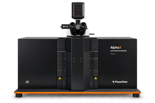
AlphaX Light Sheet Microscope
AlphaX light-sheet microscopy system

One Sample. One Platform. Multiple Scales.

From specimen overview to selected structural detail.

AlphaX combines dual-sided illumination with 2X, 5X, 10X and 20X illumination configurations on one platform. Precalibrated optical parameters coordinate transitions between imaging scales, revealing fine structures while preserving whole-specimen context.

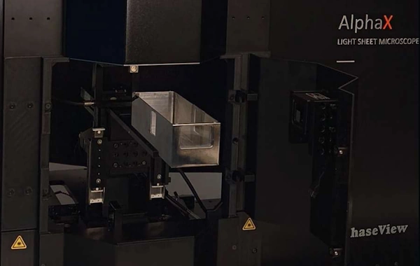
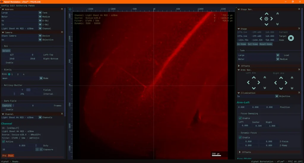
A chamber-centred design provides direct access for sample mounting and experimental interaction, while the motorized positioning mechanism remains inside the instrument. xFlow brings instrument controls, image display and workflow panels together on one screen.

MULTISCALE LIGHT-SHEET OPTICS

  • 2X Whole-specimen context
  • 5X Large-volume imaging
  • 10X Regional detail
  • 20X Fine optical sectioning

PhaseView multiscale imaging architecture

Coordinated transitions between overview and detailed imaging configurations using precalibrated optical parameters.

Thin optical sections reveal fine structures while preserving spatial context.

Dual-sided illumination

For large and optically complex specimens.

20X illumination designed for submicron theoretical optical sectioning

Approximately 0.96 µm Gaussian light-sheet FWHM at 488 nm for the 20X/0.42 NA illumination configuration. Equivalent half-waist convention: approximately 0.48 µm.

Theoretical illumination value; complete-system resolution is configuration-dependent.

Protected by intellectual-property rights.

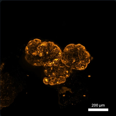
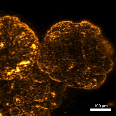
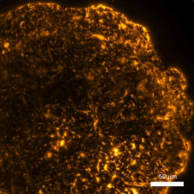
Representative organoid imaging using 5X, 10X and 20X illumination configurations

Representative organoid datasets acquired using PhaseView light-sheet microscopy. Images are maximum-intensity projections; display ranges are adjusted independently and shown using the same orange pseudo-colour. Courtesy of Dr Jacqueline Ross, University of Auckland.

Chamber-centred sample handling

Bring the Chamber to the Experiment

Direct access with the motorized positioning mechanism retained inside the instrument.

AlphaX light-sheet microscope sample chamber

Direct access
A linear loading path brings the chamber into a convenient working position.

Five-sided reach
Front, upper and side access support mounting and experimental interaction.

Separated mechanics
Helps reduce routine exposure of motorized positioning hardware to imaging media.

Visible sample
Optional viewer cameras observe the chamber and sample area.

Chamber out → Mount / Interact → Chamber in

Open for evolving experiments

The sample-bay door can remain closed for routine imaging or open when direct experimental access is required. Reduced-volume inserts and application-specific accessories can be developed around the workflow.

Open access
For custom holders and experimental accessories.

45 × 90 cm
Nominal closed-instrument footprint.

Instrument-centred software

xFlow

The instrument, the image and the workflow on one screen.

xFlow software interface for the AlphaX light-sheet microscope

Current demonstrable concepts

  • Configuration-aware instrument address
  • Micron-calibrated image display and stack navigation
  • Stage, illumination-arm and channel controls
  • Saved presets, configurable panels and multilingual interface
  • User accounts and access levels

xFlow roadmap

  • Scan sequencer
  • Processing pipeline
  • Macros and scripts
  • MCP / assistant integration
System configuration
Component Configuration
Illumination Dual-sided; 2X / 5X / 10X / 20X
Detection Vertical architecture; interchangeable objectives
Positioning Motorized XYZ; optional rotation
Chamber Large chamber, inserts and custom holders
Software xFlow
Computing Acquisition workstation + high-speed storage link

Discuss Your Research

Bring us your sample, your markers and your imaging question.

Scroll to Top