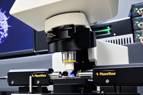
Alpha1 adds light-sheet imaging capabilities to an existing fluorescence microscope. The compact illumination unit integrates with a compatible microscope stand, detection optics and scientific camera, providing a practical path to optical sectioning and three-dimensional fluorescence imaging without replacing the complete microscope system.
Alpha1 adds light-sheet illumination to an existing fluorescence microscope.
Upgrade an Existing Microscope for Light-Sheet Imaging
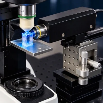
Alpha1 forms a horizontally projected light sheet through a long-working-distance illumination objective. Fluorescence is collected through the microscope detection path and recorded with a scientific camera.
The modular configuration can incorporate compatible microscope components already available in the laboratory, including the microscope stand, camera, objectives, positioning stage and sample-handling accessories.
Each Alpha1 system is configured according to the specimens, imaging medium, required field of view and existing laboratory equipment.
Sharp optical sectioning. Alpha1 forms a thin, horizontally projected light sheet for high-contrast fluorescence imaging with reduced out-of-focus background.
Integration with existing equipment. The compact illumination unit can be combined with compatible microscope stands, scientific cameras, detection objectives and positioning stages already available in the laboratory.
Open access to the sample. The accessible imaging area simplifies sample mounting, adjustment and the integration of additional experimental equipment.
Flexible excitation options. Alpha1 can be configured with compatible laser or LED illumination sources according to the fluorophores and imaging workflow.
Compact Architecture
Alpha1 uses a compact, space-efficient architecture designed for integration with compatible fluorescence microscopes. The light sheet is projected horizontally through a long-working-distance objective while the microscope’s existing detection path collects the fluorescence signal.
Accessible Sample Area
The open configuration keeps the specimen accessible during imaging. Researchers can adjust the mounting arrangement, use different sample chambers and integrate additional experimental equipment when required.
High-Contrast 3D Fluorescence Imaging
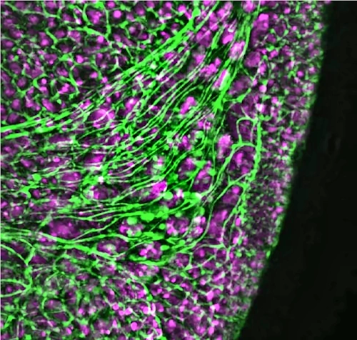
Alpha1 illuminates a thin optical section of the specimen while fluorescence is collected through the microscope’s detection objective. This reduces out-of-focus background and reveals structures within thick biological samples.
The system supports imaging of live, fixed and cleared specimens. Objectives, sample chambers and mounting arrangements are selected according to specimen dimensions, imaging medium, required field of view and spatial resolution.
Individual optical sections can be acquired as a Z-stack and subsequently visualized as a three-dimensional volume using compatible image-processing and analysis software.
QtSPIM Acquisition Software
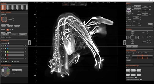
QtSPIM provides an integrated interface for Alpha1 microscope control and multidimensional image acquisition.
The software coordinates sample positioning, illumination, camera settings, fluorescence channels, Z-stacks and time-lapse acquisition within a single workflow.
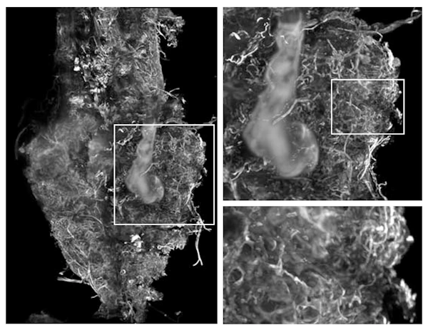
Fluorescence imaging across multiple magnifications, from specimen overview to structural detail.
From Optical Sections to 3D Data
Alpha1 supports the acquisition of individual optical sections and complete Z-stacks. Researchers can inspect the source images, navigate through the specimen and examine selected structures at different imaging scales.
Raw data are saved as 16-bit TIFF files with acquisition metadata. The resulting image stacks remain accessible for further visualization, processing and quantitative analysis using compatible open-source or commercial software.
This workflow connects microscope control, image acquisition and data review while preserving access to the original image information.