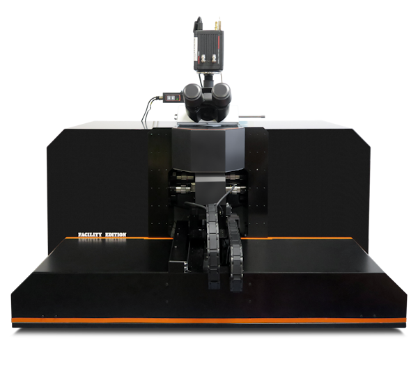
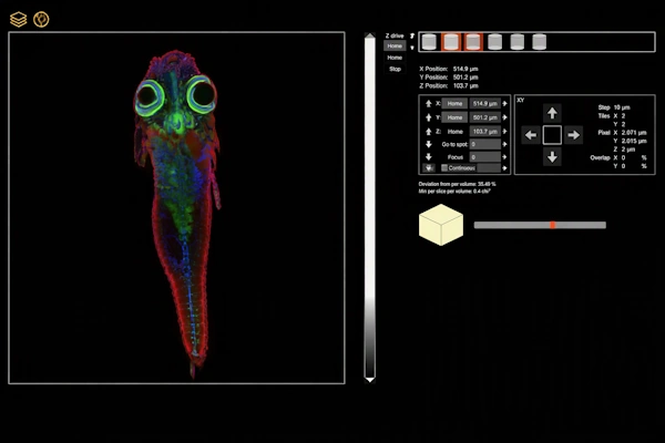
Alpha3 Facility Edition is designed for imaging facilities and laboratories working with diverse samples, users and experimental protocols. It combines the proven imaging technologies of Alpha3 with motorized optical configuration, reproducible sample handling and an integrated 3D workflow. Researchers can explore a complete specimen, select regions of interest and acquire them at higher magnification without manually rebuilding the microscope configuration.
Built for Shared Imaging Facilities
Alpha3 Facility Edition extends the Alpha3 platform with increased sample capacity, motorized objective selection and a streamlined acquisition workflow.
Researchers can move from whole-specimen overview imaging to detailed acquisition of selected regions without manually reconfiguring the microscope.
The system supports reproducible imaging of live, fixed and optically cleared biological samples across different users, specimens and experimental protocols.
From whole specimen to fine detail. Start with a rapid overview, select regions of interest and acquire them at higher magnification without manually reconfiguring the microscope.
More samples in a single experiment. A larger chamber, dedicated holders and motorized positioning support reproducible acquisition from multiple specimens or regions of interest.
Consistent image quality across the field. Dual-sided illumination, active focus sweeping and optimized illumination optics provide thin, uniform optical sectioning for large and complex samples.
A streamlined 3D imaging workflow. Rapid preview, interactive volume definition and integrated microscope control simplify setup and reduce repeated manual adjustments.
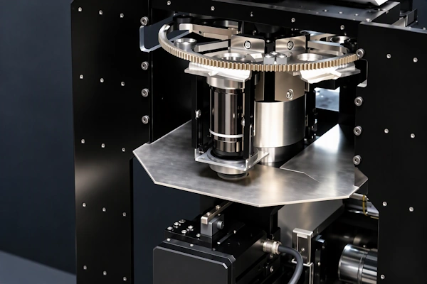
Motorized objective selection for automated multiscale imaging.
Proven Alpha3 Imaging Technologies
Alpha3 Facility Edition retains the core illumination technologies of the Alpha3 platform and combines them with motorized optical adjustment for automated imaging workflows.
Dual-Sided Illumination
Two opposing illumination units improve light coverage and help reduce shadowing in absorbing or scattering biological samples.
High Illumination NA
High-numerical-aperture illumination produces a thin light sheet for improved optical sectioning and finer structural detail.
Real-Time Focus Sweeping
The optimal illumination region moves across the camera field to maintain more uniform sharpness and light-sheet quality across the image.
High Lateral Illumination NA
Illumination from multiple lateral angles improves intensity homogeneity and helps reduce directional illumination artefacts.
Automated Multiscale Imaging
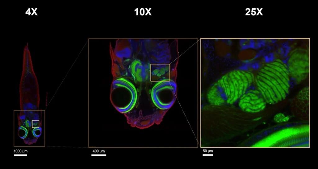
Alpha3 Facility Edition supports a complete multiscale workflow, from rapid whole-specimen overview imaging to detailed acquisition of selected regions of interest.
When the magnification changes, motorized illumination and detection objective turrets automatically select the appropriate objectives. The system then adjusts the optical configuration for the new imaging scale.
This automated workflow allows researchers to locate structures within a large specimen and investigate selected areas in greater detail without manually reconfiguring the microscope.
Automated multiscale workflow from whole-specimen overview to detailed imaging of selected regions.
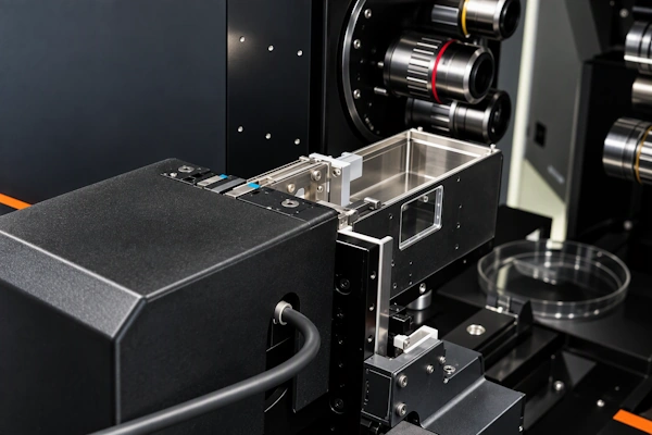
Multi-sample holder for reproducible automated imaging.
Reproducible Multi-Sample Imaging
Alpha3 Facility Edition supports convenient loading and reproducible positioning of individual or multiple specimens. Dedicated holders accommodate samples of different sizes, from small model organisms to cleared tissues and whole organs.
Motorized sample positioning and acquisition presets allow multiple specimens or regions of interest to be imaged within the same experiment, reducing repeated manual setup.
Optimized Workflow with LINDA
Alpha3 Facility Edition guides the imaging workflow from sample loading and rapid preview to acquisition-volume definition and final 3D imaging.
Fast Sample Preview
Low-magnification imaging provides a rapid overview of the specimen and helps identify relevant structures before full acquisition.
Interactive 3D Volume Definition
LINDA progressively displays the preview volume and allows single or multiple regions of interest to be selected directly in 3D.
Automated Acquisition Setup
The system configures the required objectives, imaging parameters and acquisition positions for the selected volumes.
Volume Acquisition and Data Export
Defined regions are acquired automatically, with image data and metadata available for further visualization, processing and analysis.
LINDA Slice View for specimen navigation and acquisition-volume setup.
Discuss Your Imaging Requirements
Tell us about your specimens, preparation methods, imaging protocols and facility workflow. We can discuss an Alpha3 Facility Edition configuration for automated multiscale and multi-sample imaging.
The system can be configured to support your current experiments while remaining adaptable to new samples, users and research requirements.